
Published by: Nuru
Published date: 17 Jan 2022
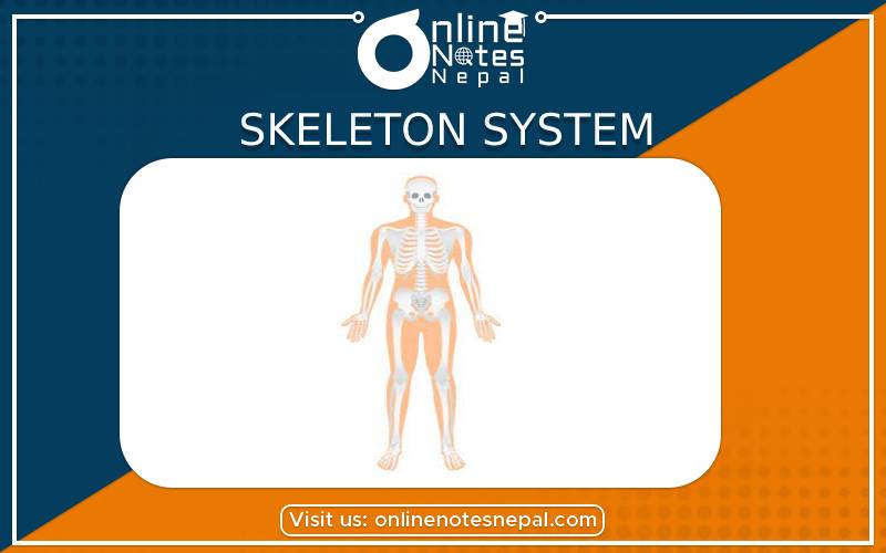
Skeleton system is a supporting framework of the body made by the combination of various bones. There are 206 bones in our body.
Functions of bones are:
Bones are divided into two groups:
Functions of the skeletal system are as follows:
Skull:
The skull consists of the bones of the head and it is divided into cranium and facial bones. Altogether, there are twenty nine bones in the skull.
Cranium: The cranium is a large bony box and is composed of eight flat bones. One bone of the cranium interlocking with the edge of another bone by saw-like edges is known as suture. The bone of cranium are as follows:
Frontal bone:
The frontal bone is one in number and forms the forehead or the front of the cranium.
Parietal bone: The parietal bones are two in number and form anterior sides and the roof of the skull.
Temporal bone: The temporal bones ate two in number and lie on each side of the head and form temples and complete the side-walls of the skull.
Occipital bone: The occipital bone is one in number and forms the back of the head and the posterior base of the skull.
Sphenoid bone: The sphenoid is one in number and it is bat shaped bone.
Ethmoid bone: The ethmoid is one in number and lies in front of the sphenoid.
Facial bones:
The facial part of the skull is formed of fourteen bones. All of them except the lower jaw are immovable. The bones of the facial parts are as follows:
Mandible or lower jaw bone: Mandible bone is only movable bone of the skull having 16 sockets for the lower set of the teeth on its upper edge.
Maxillae or upper law bones: Maxillae bone forms a portion of the roof of the mouth, lateral walls of the nasal cavity and part of the floor of the orbital cavities.
Palatine bone: Palatine bone are 'L' shaped and forms the upper part of the palate or roof of the mouth cavity.
Zygomatic bones: There are two zygomatic bones which form the prominences of the cheeks and part of the floor and lateral wall of the orbit eyes.
Vomer bone: It is a thin and flat single bone that extends upwards and forms the part of the middle partition in the nose and divides the two nostrils of the nose.
Nasal bone: Two small flat bones that form the bridge of the nose. The lower part of the nose is formed of cartilage.
Inferior nasal conchae: The two conchae are bones of the nose, one on each chamber of it.
Lacrimal bone: These are two small lacrimal bones in inner wall of the orbit of the eyes. Lacrimal bone contains the passage for tears from the eyes to the nose.
Ear ossicles:
There are three bones in the middle ear which are called ear ossicles. It consists of two malleus, two incus and two stapes in the ears. The stapes are the smallest bones of the body.
Hyoid:
Hyoid is a ‘U’ shaped single bone and it supports our tongue. It is located in the neck region below the lower jaw.
Bones of the trunk
Trunk consists of bones of the vertebral column and the thorax.
Vertebral column:
The vertebral column supports the head, helps in upright posture and locomotion. The individual bones of the vertebral column are known as vertebrae. Each vertebra is provided with the canal called neurocoel.
Cervical vertebrae: The first two cervical vertebrae are different from all other vertebrae. The first cervical vertebra is called atlas. It supports the globe-like head. The second cervical vertebra is called axis. It helps in movement of head from side to side.
Thoracic vertebrae: Thoracic vertebrae ate 12 in number and form the upper part of the back.
Lumber vertebrae: Lumber vertebrae are 5 in number and form the lower part of the back.
Sacral vertebrae or sacrum: In infants sacral vertebrae consist of 5 separate bones but in adult they are found fused to form a large wedge-shaped sacrum.
Coccygeal vertebrae or coccyx: In infant, coccygeal vertebrae consist of 4 separate bones but in adult they are fused to form a very small triangular bone.
Thoracic bones:
There are 12 pairs of semi-circular bones present in the thorax called ribs and a long and broad bone called sternum. The spaces between the ribs are called intercostal spaces which are occupied by intercostal muscles.
True ribs: The first seven pairs of ribs are directly attached to the sternum by means of cartilage called true ribs.
False ribs: The 3 pairs of ribs are attached to that of the seventh pair of ribs and not to sternum directly are called false ribs.
Floating ribs: The last true ribs which are free and do not even reach the sternum are called floating ribs.
They are divided into two parts
1. Shoulder gridle:
The bones in our hand are:
Humerus: The long bone extending from shoulder to elbow is called humerus.
Radius and Ulna: The part between elbow and wrist consists of two bones called radius and ulna. The ulna lies towards the little finger and the radius lies towards the thumb.
Carpals: There are 8 carpals in the wrist. They lie as two rows, four in each row.
Metacarpals: Each hand consists of five metacarpals. They are attached to carpals at one side and two fingers at the other side.
Phalange: The five fingers consist of 14 pieces of bones called phalanges. The thumb consists of two bones and the remaining four fingers consist of three bones each.
Pelvic girdle: The two bones connected to the thigh bones and vertebrae together are called pelvic gridle.It helps to protect the organ like stomach, urinary bladder and intestine.
The bones at legs are:
Femur: The largest and the strongest bone of the body is femur. It is attached to the tibia and fibula bone at the lower part.
Fibula and Tibia: They are two long bones below the knee. One is fibula which is thin and weak. It does not the support the load of the body. The other is tibia which is thick and strong. It supports the load of the body.
Tarsals and Metatarsals: There are 7 bones in the tarsals whereas metatarsals are made up of five bones. Tarsals are round and are arranged in three rows. Metatarsals are straight and long.
Phalanges: The toes consist of 14 pieces of bones as in the hands. The first toe consists of two bones and the rest consist of three bones each.
The joining of two or more bone is called joint. There are four types of joint in a body. They are as follows: